Abstract
Purpose: To describe the clinical presentation and novel anatomical features of a patient with chronic central serous chorioretinopathy (CSCR) complicated by retinal neovascularization (RNV).
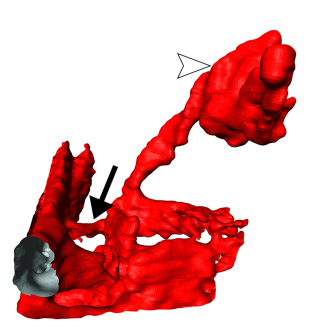
Observations: A 48 year-old patient with a long-standing history of bilateral CSCR presented to our clinic complaining about a sudden onset of tiny floaters. Multimodal imaging including fundus autofluorescence (FAF), fundus fluorescein (FA) and ICG angiography (ICG) and spectral domain optical coherence tomography (SD-OCT) confirmed the diagnosis of CSCR and revealed a pre-retinal neovascularization and concurring vitreous hemorrhage. Swept source OCT angiography (OCTA) and 3D reconstruction virtual reality determined the retinal origin of the neovascularization. Follow-up examination revealed clearing of the vitreous hemorrhage and spontaneous obliteration of the RNV without any treatment three months following the initial presentation.
Conclusion and importance: To the best of our knowledge, this is the first report of a RNV associated with CSCR which was determined by three-dimensional (3D) OCTA reconstruction.
Gruber M, Wolf J, Stahl A, et al. Novel insights into retinal neovascularization secondary to central serous chorioretinopathy using 3D optical coherence tomography angiography. American journal of ophthalmology case reports 2020;18:100609.