Maloca PM, Tufail A, Hasler PW, Rothenbuehler S, Egan C, Ramos de Carvalho JE, Spaide RF. Acta Ophthalmol. 2017 Dec 14. doi: 10.1111/aos.13637.
PURPOSE. To demonstrate the feasibility of three dimensional (3D) printing of choroidal vessels and pigmented choroidal tumors derived from volume optical coherence tomography data.
METHODS. 1050 nm swept source optical coherence tomography (SSOCT) volume data were acquired with DRI OCT Triton (Topcon, Tokyo, Japan). For the 3D printing, OCT volumes were freed from speckle noise using a recently developed 3D denoiser. Three-dimensional information of the processed choroid (choroidopsy) was saved as obj-file for sealing gaps in the mesh or remove obvious artifacts what resulted in a 3D printable OCT model. Several printing materials and model sizes were tested for durability and for depicting the microarchitecture of choroidal vessels and tumors.
RESULTS. The proposed method allowed 3D printing of choroidal vessels of Haller’s and Sattler’s layer. Polyamides and transparent resin showed excellent material properties with high resistance and possibilities for further processing with gold. The polymer gypsum powder coating method showed good usability in two-colour printing but was too fragile.
CONCLUSION. The developed method provided a potentially useful tool for 3D printing and 3D analysis of OCT.

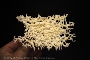
First printed choroidal 3D model in 2011 by Cyrill Gyger using selective laser sintering with natural white polyamide shows a healthy, dense central choroidal vessel network of the macula. The gap to the left depicts the area occupied by the optic disc (model size 120 mm x 75 mm x 8 mm obtained from 1050 nm OCT data). © 2017 OCTlab University of Basel, Switzerland. All rights reserved.

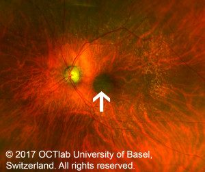
Retinal fundus imaging of a central, dark pigmented choroidal tumor (arrow). Only a few choridal vessels are preserved which are visible as fine, red lines. © 2017 OCTlab University of Basel, Switzerland. All rights reserved.

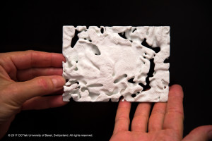
3D print of the same macula tumor of the previous image. The tumor shows some openings which are caused by the preserved choroidal vessels. © 2017 OCTlab University of Basel, Switzerland. All rights reserved.

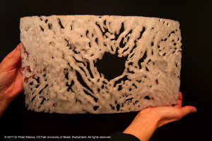
Fused deposition modeling (FDM) printed model using transparent polycarbonate (back side view). The largest 3D print model printed to date depicts the dense choroidal vascular arch around a healthy optic nerve which is not depicted (Circle of Zinn or Haller, model size 210 mm x 390 mm x 23 mm). © 2017 OCTlab University of Basel, Switzerland. All rights reserved.
